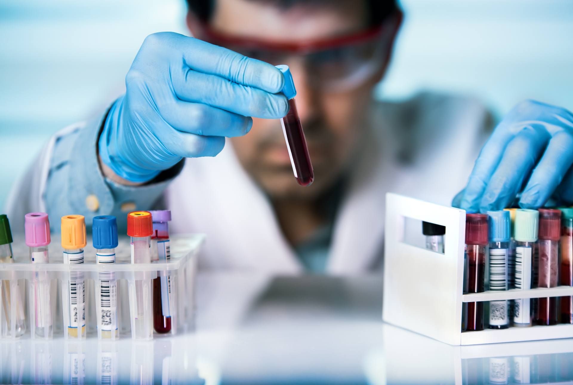
 An image is reconstructed from the position of the detected photons as they depart the body creating a picture of biochemical exercise occurring on the diseae web site and in the entire body. PET scanning has been a clinically valuable method for each research and affected person administration since the Eighties. However, zenwriting.Net it's only been inside the last 20 years that the Centers for Medicare & Medicaid Services (CMS) has reimbursed for clinical PET research, making them widely available in the US. Newer technology combines PET and CT into one scanner, known as PET/CT. PET/CT shows explicit promise within the diagnosis and therapy of lung cancer, evaluating epilepsy, Alzheimer's disease and coronary artery illness.
What is a PET scan?
Remember to keep away from chewing gum or laboratorio analises Clinicas Veterinaria sucking on exhausting sweet, cough drops, or mints. If you’re receiving a PET/CT scan, additional tracer will be wanted. This may be harmful to individuals who have kidney illness or who've elevated creatinine ranges from medications they’re already taking. People who are allergic to iodine, aspartame, or saccharin ought to let their doctor know. The dangers of the test are also minimal compared to how useful the outcomes could be in diagnosing critical medical conditions. In the United States, around 2 million PET scans are carried out every year, in accordance with Berkley Lab. However, it will depend on the company and coverage you buy.
Mediante el empleo de esta técnica podemos llegar a advertir el agravamiento de la patología y también intentar eludir probables descompensaciones del corazón, que acarrean el coherente empeoramiento en la calidad de vida de nuestros compañeros.
It may also be used to judge whether or not there is harm to heart muscle and weakened pumping perform from a heart assault. In addition, you might even see cardiac ultrasound known as "Transthoracic" or Transesophageal" echocardiography. Transthoracic Echocardiography (TTE) is when a cardiac ultrasound is carried out on the patient’s chest. TTE is the commonest cardiac ultrasound software and is non-invasive. Transesophageal Echocardiography (TEE) is a more specialised cardiac ultrasound with a particular probe that is inserted into the patient’s esophagus.
var rs = document.createElement('script');
During the echo check, you’ll be asked to placed on a hospital gown. You’ll lie on an examination desk, and a sonographer or ultrasound tech will put some gel on the end of an ultrasound wand and move it alongside your chest. The gel might be a little cold, however otherwise you should not really feel any major discomfort in the course of the take a look at. They might also order an echocardiogram if something irregular, like a coronary heart murmur, is detected during an examination. It can help them see if there’s been injury to the center muscle, or an issue with a valve, Dr. Miyasaka says. Since the bottom pressures in the coronary heart is the best atrium, the primary echo signal you will notice of cardiac tamponade is correct atrial systolic collapse. Point of Care Ultrasound can provide a more definitive diagnoses of pericardial effusion and cardiac tamponade.
Conditions
A transesophageal echocardiogram entails an area anesthetic in your throat. Your supplier will introduce a flexing tube down your esophagus to your stomach. This device then information pictures of your coronary heart to an exterior monitor that your supplier will view. This take a look at entails a sonographer using a tool like an ultrasound machine to report live images of your coronary heart and its valves. If they need a better have a look at how your valves are functioning, medical doctors might order a transesophageal echocardiogram.
The speed and incline of the treadmill or bike will increase every couple of minutes until you attain the target heart rate, or till you would possibly be too drained. Based on the pictures and observations made throughout an echocardiogram, your healthcare provider can decide the presence and severity of sure conditions. They can use this info to suggest efficient therapy. There are several varieties of echocardiogram tests and a number of other causes for why healthcare suppliers use them. The take a look at has been round for an extended time, but the quality of the pictures that docs can get with it continues to enhance. An echo will present whether or not the four heart valves are opening and closing correctly as the center pumps to direct adequate blood flow in the right direction.
Why would my doctor do an echocardiogram?
A system called a transducer will be placed on your chest over your coronary heart. The transducer sends ultrasound waves via your chest toward your heart. A laptop interprets the sound waves as they bounce again to the transducer. Often, a technician does some or all the take a look at, however a doctor, normally a heart specialist, will take a glance at your coronary heart photographs when you are having your echo. They could want to adjust the transducer (the handheld device used) to visualise extra views, if needed.
Are you getting health care you don't need?
The stress echo often takes about 20 to 30 minutes however can vary relying on how lengthy you exercise or how lengthy it takes the medicine to boost your heart price. In some situations, a TEE could additionally be ordered after echo results are reviewed, significantly if your doctors are involved that you've a coronary heart downside that was not detected. A TEE looks on the heart by putting an ultrasound device inside your esophagus, as an alternative of outdoor your chest. There are professionals and cons to each checks, and essentially the most important difference is that TEE is invasive and requires sedation. Because a transesophageal echo is an invasive check, you may be given a sedative and oxygen during the take a look at. Your throat will be numbed and your provider will insert a flexible tube down your throat. The sound waves that create the picture of your heart are released from a transducer on the end of the tube.


